Tuesday, August 21, 2012

Summer Internship at a Biorepository

Matthew Masiello spent the summer interning with our tissue bank at Windber Research Institute. He will be pursuing a degree in International Relations from American University in Washington, DC this fall. 


By Matthew Masiello
As high school was coming to an end, I, as well as a multitude of my peers was on the look for summer jobs, searching for positions that would sustain us throughout the summer and hopefully be able to support maybe a fraction of the total cost of the college that was approaching in the fall. It just so happened that as my friends found themselves working in restaurants, manual labor, or totally bypassing the opportunity to work during the summer before their new scholarly life, I found myself walking into the doors at Windber Research Institute ready to begin my internship within the tissue bank.

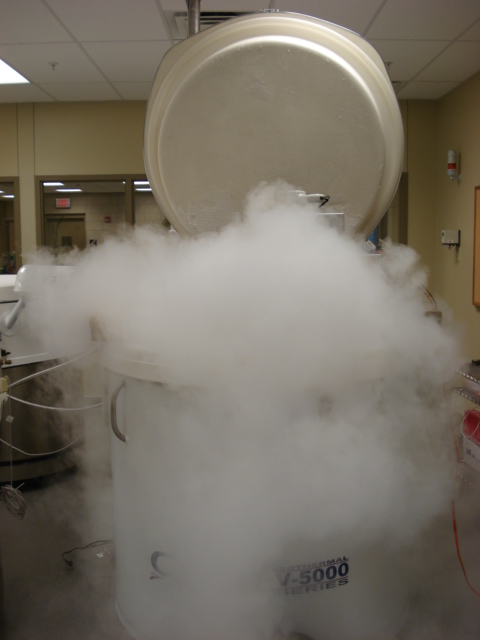
The tissue bank at WRI provides the optimal isothermal freezer space for storing blood, tissue, and DNA samples. Over the past decade the tissue bank has gone from great to greater and to maintain such levels of efficiency and reliability certain procedures are instilled to monitor the isothermal freezers and their samples inside. So as an intern, my assignment was to perform a Quality Assurance project on the mass amount of DNA samples that the freezers contain, meaning that I was to sort through boxes of DNA, confirming specific sample locations and identification with the extensive software program that accounts for all samples. Up front, it was a task I was ready to embrace, however, I do suppose every job comes with its own fair share of difficulties and mine just happened to be enduring cold hands/fingertips due to handling samples kept at -80° Celsius or less and squinting through dry eyes because of my proximity to the dry ice that I used daily to perform my work. However, through determination and hand-warmers I accomplished my task and felt as though I was contributing to an already efficient system.

Another task I had the opportunity to explore was blood processing.  This procedure involves centrifuging, labeling, aliquoting and scanning the samples into a sample management software program.  Efficiency and organization were in place, common themes at the tissue bank. Honestly, this process was a humbling experience at the beginning of my time at WRI.  It would have never come across my mind if I had worked at any other sort of job that my hands resemble the San Andreas Fault when it came to using the pipette to draw from the sample tube. Truly, only at a tissue bank do you find what your actual natural abilities are. Thankfully, at the same rate I found myself truly enjoying the process as I was becoming better at handling the pipette and more and more efficient with the task at hand.

My internship at WRI’s tissue bank was not only scientifically satisfying, but also prepped me for what I should expect with any sort of occupation I might find myself in the future. Efficient management and clear-cut organization are key factors to any system that strives to be the best and both are clearly exemplified at the tissue bank, and I will guarantee that I remember my time here and use the skills and styles that I developed and apply them to any sort of work I find myself conducting in the approaching years.

Friday, June 22, 2012

When You Need to Re-evaluate Tissue pathology Data

When we receive pathology report for data entry, we usually review this to determine it matches the slide available before the information goes through the data entry step. If we therefore find that an  OCT.specimen has a diagnosis that does not correlate to the received participant information (for example we have tumor size 1.0 cm or greater; while OCT diagnosis – no tumor seen) the OCT block will be tracked and retrieved for re-evaluation. Deeper sections will becut for a second review. A record is kept of all the samples that we consider for deeper sections. This record shows the information received, along with the diagnosis of the first evaluation of the H&E slide. After the second evaluation is performed, the information is added to the record.
This re-evaluation process, as we call it, has been very helpful. The deeper cuts, in some cases, have revealed tumors making these samples suitable for certain research protocols thus increasing the number of material in our tissue bank that can be utilized for research.  This is always something that the research scientsist are happy about!

Thursday, May 17, 2012

Researching Our Biospecimens

The tissue bank at the Windber Research Institute focuses on collecting and storing well annotated and high quality tissue and blood samples.  In order to demonstrate that the samples are of the highest quality, we do biospecimen research on samples in house to test different processing methods or storage conditions and determine which are best.  Pre-analytical variables that could affect downstream research processes that utilize DNA or RNA are studied in ongoing projects at our tissue bank.  High DNA/RNA concentration and good yield, integrity and purity are important in all experiments. 
            One such experiment is our biospecimen research project where we looked at the integrity and purity of RNA that was isolated from breast tissue collected from reductive mammoplasties.  Tissue specimens were processed by flash freezing or embedding in optimal cutting temperature (OCT) medium and either immediately stored at -80°C or kept at room temperature over varying time intervals.  RNA isolation was performed on all samples and the Nanodrop spectrophotometer was used to determine the concentration and purity of the RNA.  RNA integrity number was also determined using the Agilent Bioanaylzer.  Since laser capture microdissection is sometimes an integral part of tissue analysis, we also determined its effect on RNA integrity.  Quantitative Real-Time PCR was done for comparative measurement of the expression of four house keeping genes: B-actin, glyceraldehyde-3-phosphate dehydrogenase, cyclophilin A and porphobilinogen deaminase.  Results of this study can be viewed on the additional poster.

Wednesday, May 2, 2012

A Cut above the Rest

Laser Microdissection is an intermediate step in the preparation of tissue specimens for research.  This service is provided by our tissue bank and gives researchers the ability to obtain macromolecules (DNA, RNA and protein) from specific cell types of a tissue specimen.  There are three Microdissection Microscopes currently in the market- Arcturus Pixcell, Leica Lasermicrodissection ASLMD and the Zeiss PALM.  We currently use the Leica ASLMD system.

Our primary focus in Windber is the study of breast tissue which is highly heterogeneous with numerous cell types within the primary tumor and the surrounding tissue.  Laser microdissection has allowed us to isolate pure samples from both invasive and in situ tumors (with and without involvement within invasive tumors), as well as the microenvironment surrounding the tumors that can then be used for downstream analyses.

We have used Laser Microdissected tissue samples for a variety of protocols requiring the preparation of homogenous isolates of DNA, RNA and protein.  Because the quality of RNA extracted from tissues can be affected by sample preparation and processing, we have fine-tuned our procedures for processing these samples.  Our protocols include careful cleaning of all equipment with RNAse inhibitors, use of special stains with lower water content, and rapidly performing the isolation process.  The time to process each sample is kept to less than 30 minutes from the time the specimen is sectioned through staining, microdissection and placement into the cell lysis buffer.  Below are images of a ductal carcinoma in situ (DCIS) from which RNA was isolated from the Microdissected cells.  The image labeled (A) is an Hematoxylin and Eosin Stain; (B) is a representative section in the same area stained with the RNA staining procedure and image (C) is the DCIS after Microdissection.

We have published details of our protocols in the chapter titled- “Laser Microdissection for Gene Expression Profiling” in the book “Laser Capture Microdissection Methods and Protocols Series: Methods in Molecular Biology”, Vol 755, Second Edition, Humana Press. 

A

B


C


Thursday, April 19, 2012

WRI in Bloom

 As I think about Windber Research Institute (WRI) in bloom this spring, I am reminded of the renewal and regrowth we experienced in 2005.  As our staff and research expanded, we outgrew the facility originally built for ten to fifteen people.


St. Mary's Church
In August 2005, Windber Research Institute moved to the location on the former site of St. Mary's Hungarian Roman Catholic Church. In an effort to honor the support from the Windber community, the arch of St. Mary's Church was preserved and incorporated into the design of the new facility.

Just as WRI has grown over the years, the tissue bank continues to blossom as well.  We have gone from our first batch of samples in 2001 when electronic records were scant, to our current status where electronic records abound…shipping manifests, databases, checklists, reports, automated monitoring, QA… we have it covered.  It is amazing to look back at where we were in our infancy and see how we have grown into a mature biorepository in 2012.  

Windber Research Institute
Just as spring brings new growth after the winter hibernation, exciting opportunities are upon us!  A most encouraging development is our new partnership with the U.S. Military Cancer Institute to handle their biobanking needs.  We recently expanded our tissue bank adding additional space and freezers to accommodate frozen tissue specimens from the U.S. Military Cancer Institute’s research program.  This contract serves as a vote of confidence in our leading edge facility and biobanking team and we are proud to be working with them.

Friday, April 6, 2012

Maintaining the Freezers 24/7


Preserving the integrity of our precious samples is a top priority in our tissue bank.  We recognize that if it were not for the generosity of the patients, this great resource would not exist.  To this end, daily checks are performed to ensure that everything is operating smoothly and within acceptable limits.

WRI’s Tissue Bank consists of ten liquid nitrogen freezers and five negative 80 freezers which are continuously monitored by an automated system. If a temperature falls out of range, the system will call staff from a designated call list so the necessary action can be taken.

In addition to the automated system, we also perform manual checks as a backup method.  Every day, two staff members perform AM and PM checks of the tissue bank.  The checklist includes room temperatures, freezer temperatures, nitrogen levels, oxygen levels and verification that freezers are locked.  During non-business hours, security staff walks through the facility and performs the same checks every four hours.

All checklists are double checked at the beginning of every week to look for possible trending patterns.  This process can help to give us a heads up of a possible problem in the future.  One such occasion was when we noticed liquid nitrogen levels rising instead of falling on one of the freezers (this parameter is not recorded on the automated system).  We did some troubleshooting and ended up needing to do a total thaw of the freezer to clear out an obstruction that was leading to the erroneous readings.

Maintaining the tissue bank is a 24 hour 7 day a week job, and every day many steps are taken to ensure the quality and integrity of our samples remain high.


Wednesday, March 21, 2012

Combing Through Samples to Eliminate Knots


Here at WRI, we have a growing inventory of Clinical Breast Care Project (CBCP) blood and tissue samples totaling 80,000 aliquots.  We have been collecting and storing samples since 2001.  Every sample donated is a valuable asset for our biomedical research activities.  We manage our inventory using quality standards so that we can provide high quality specimens to our research scientists.  Therefore our aim is to reduce errors to the barest minimum (as is humanly possible).   
To ensure these goals are met, we perform regular Sample Quality Assurance (SQA). One of our SQA was performed on CBCP samples collected prior to December 2007.  We predicted there would be some errors made in the early days of sample collection especially since we have modified our Standard Operating Procedures (SOP) over the years.  This SQA was the first that represented a time frame of this magnitude.
The project was initiated in August 2010 and completed in December 2011.  Boxes were physically pulled from the freezers and compared to sample records in our electronic databases.  It was not practical to check every individual sample so we formulated a plan to check 20% of blood samples per box and 100% of tissue samples per box.  Sample discrepancies were corrected and logged onto our Quality Assurance tracking database.  Paper and electronic records were kept during the SQA project to monitor progress and keep track of corrections.
The SQA project allowed us to check 19.7% of our total CBCP inventory.  We observed an error rate of 2.5%.The most common errors were from hand written information on sample tubes. We observed during this SQA exercise that errors were more common during the earlier years when we did not utilize a bar coding system. 
 We have since implemented a bar-coding system which has decreased data entry time and errors.  We also continually update and improve our SOP’s for sample collection, processing, and storage.  All inventory errors have so far been corrected.